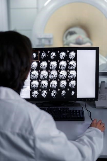
Examination of Cranial Nerves
The examination of cranial nerves is an important part of a neurological examination. It helps to identify any dysfunction or damage to the cranial nerves‚ which can be caused by a variety of conditions‚ such as stroke‚ tumor‚ or trauma. This examination involves assessing the function of each cranial nerve‚ using a variety of tests and observations. The results of the cranial nerve examination can help to pinpoint the location and nature of the neurological problem‚ aiding in diagnosis and treatment.
Introduction
The human nervous system is a complex network responsible for controlling and coordinating all bodily functions. A crucial component of this intricate system are the cranial nerves‚ twelve pairs of nerves that emerge directly from the brain. These nerves play a vital role in various sensory and motor functions‚ including vision‚ hearing‚ taste‚ smell‚ facial expression‚ and swallowing. Examining the cranial nerves is a fundamental aspect of a neurological assessment‚ providing valuable insights into the health and functionality of the nervous system.
A thorough examination of cranial nerves can help detect and localize neurological dysfunction. This assessment is crucial for identifying a variety of neurological conditions‚ such as stroke‚ brain tumors‚ multiple sclerosis‚ and peripheral nerve disorders. The examination involves systematically assessing the function of each cranial nerve through a series of tests and observations‚ allowing healthcare professionals to pinpoint the specific nerve or area of the nervous system affected.
Anatomy

The cranial nerves are a unique part of the peripheral nervous system‚ originating from nuclei within the brain and exiting the skull through foramina and fissures. Their numerical order‚ from I to XII‚ is determined by their exit location from the skull‚ moving from rostral to caudal. While most cranial nerves emerge from the brainstem‚ the olfactory (I) and optic (II) nerves originate from the forebrain‚ and the accessory (XI) nerve has a nucleus in the spinal cord.
Each cranial nerve has a specific function‚ some carrying sensory information from the head and neck‚ others controlling motor functions of facial muscles‚ and some responsible for both sensory and motor functions. This complex system of nerves allows for a wide range of sensory experiences‚ motor control‚ and autonomic functions crucial for daily life. Understanding the anatomy of each cranial nerve is essential for interpreting the findings of a neurological examination and accurately diagnosing any potential pathologies.
Clinical Significance
The examination of cranial nerves holds significant clinical importance as it serves as a crucial tool for neurologists and other healthcare professionals in diagnosing and evaluating a wide range of neurological conditions. By assessing the function of each cranial nerve‚ clinicians can identify potential pathologies affecting the brain‚ brainstem‚ or peripheral nerves. The findings from a cranial nerve examination can help localize the site of the lesion‚ determine the severity of the neurological impairment‚ and guide further investigations and treatment strategies.
Furthermore‚ the examination of cranial nerves can provide valuable insights into the progression of neurological diseases‚ monitor the effectiveness of treatment interventions‚ and identify potential complications. For instance‚ a change in pupillary response or facial muscle weakness during a follow-up examination may indicate a worsening of a neurological condition or a new development. Therefore‚ the cranial nerve examination plays a vital role in the comprehensive assessment and management of neurological disorders.
Cranial Nerve Examination
The cranial nerve examination is a systematic assessment of the twelve pairs of cranial nerves‚ which originate from the brain and control various sensory and motor functions of the head and neck. This examination involves a series of specific tests and observations designed to evaluate the integrity and function of each nerve. It is typically performed as part of a comprehensive neurological examination to identify any abnormalities or deficits that may indicate underlying neurological disorders.
The cranial nerve examination typically begins with a thorough history taking to gather information about the patient’s symptoms and medical history. This is followed by a physical examination that includes observing the patient’s appearance‚ gait‚ and posture. The examiner then proceeds to test each cranial nerve individually‚ using standardized techniques to assess sensory functions‚ motor functions‚ and reflexes. The findings from the examination are carefully documented and interpreted to determine the presence and nature of any neurological impairment.
Preparation
Before embarking on the cranial nerve examination‚ adequate preparation is crucial to ensure a smooth and effective assessment. This involves creating a conducive environment‚ gathering necessary materials‚ and ensuring the patient is comfortable and understands the procedure.
A well-lit examination room is essential for clear visualization during the examination. A quiet and private setting can minimize distractions and allow for focused testing. Gather the necessary equipment‚ including a penlight‚ ophthalmoscope‚ tuning fork‚ cotton swab‚ and a set of scent vials.
Prior to the examination‚ introduce yourself to the patient‚ verify their identity‚ and explain the purpose of the examination. Obtain informed consent from the patient‚ ensuring they understand the procedure and any potential risks or discomforts involved. Finally‚ ensure the patient is comfortably positioned in a sitting position with their head at a level that allows for clear observation and testing of the cranial nerves.
Examination of Cranial Nerves I-VI
The examination of cranial nerves I through VI encompasses the assessment of smell‚ vision‚ eye movements‚ and pupillary responses. Each nerve is evaluated systematically‚ ensuring a thorough understanding of its function and any potential abnormalities.
Cranial Nerve I‚ the olfactory nerve‚ is tested by presenting the patient with a series of non-pungent odorous substances‚ asking them to identify the scent. Cranial Nerve II‚ the optic nerve‚ is examined through visual acuity testing‚ assessment of visual fields‚ and fundoscopic examination to inspect the retina.
Cranial Nerves III‚ IV‚ and VI‚ the oculomotor‚ trochlear‚ and abducens nerves‚ control eye movements. These nerves are evaluated by observing the patient’s eye movements in all directions‚ assessing for any limitations‚ asymmetry‚ or involuntary movements. Pupillary responses are assessed by shining a light into the patient’s eye and observing the constriction of both the ipsilateral and contralateral pupils.
Cranial Nerve V‚ the trigeminal nerve‚ controls facial sensation and mastication. The sensory component is evaluated by testing light touch‚ pain‚ and temperature sensation on the face‚ while the motor component is assessed by observing the patient’s jaw movements‚ including clenching and lateral movements.
Examination of Cranial Nerves VII-XII
The examination of cranial nerves VII through XII delves into the assessment of facial expressions‚ hearing‚ taste‚ swallowing‚ and tongue movements. This part of the examination requires meticulous observation and specific tests to assess the integrity of these crucial nerves.
Cranial Nerve VII‚ the facial nerve‚ is responsible for facial expressions and taste sensation. It is evaluated by observing the patient’s facial symmetry at rest and during various expressions‚ such as smiling‚ frowning‚ and raising eyebrows. Taste is tested by applying a sweet‚ salty‚ sour‚ or bitter substance to the anterior two-thirds of the tongue.
Cranial Nerve VIII‚ the vestibulocochlear nerve‚ controls hearing and balance. Hearing is assessed by using a tuning fork or audiometry‚ while balance is evaluated through various tests‚ such as the Romberg test and the finger-to-nose test.
Cranial Nerves IX and X‚ the glossopharyngeal and vagus nerves‚ are responsible for swallowing‚ taste‚ and speech. These nerves are evaluated by observing the patient’s swallowing ability‚ palatal movement‚ and gag reflex. Voice quality and articulation are also assessed.
Cranial Nerve XI‚ the accessory nerve‚ controls head and shoulder movements. It is assessed by observing the patient’s ability to shrug their shoulders against resistance and turn their head against resistance.
Cranial Nerve XII‚ the hypoglossal nerve‚ controls tongue movements. It is evaluated by observing the patient’s tongue movements‚ including protrusion‚ lateral movement‚ and articulation.
Documentation and Interpretation
Accurate documentation of the cranial nerve examination is crucial for effective communication and patient care. The findings should be recorded clearly and concisely‚ including details about each nerve’s function and any observed abnormalities. This documentation serves as a valuable record for the patient’s medical history and facilitates communication between healthcare professionals.
Interpretation of the examination results requires careful consideration of the patient’s clinical presentation‚ medical history‚ and other diagnostic findings. For example‚ unilateral weakness of the facial muscles could suggest a peripheral nerve lesion‚ while bilateral weakness might point to a central nervous system disorder. The specific pattern of cranial nerve involvement can provide valuable clues to the underlying pathology.
A thorough understanding of the anatomy‚ function‚ and potential pathologies associated with each cranial nerve is essential for accurate interpretation. Understanding the relationship between cranial nerve dysfunction and the affected brain regions is crucial for formulating appropriate diagnostic and treatment plans.
The cranial nerve examination is a valuable tool for assessing neurological function and identifying potential underlying conditions. Accurate documentation and careful interpretation of the findings are essential for providing optimal patient care.
Clinical Applications
The examination of cranial nerves has a wide range of clinical applications‚ playing a vital role in the diagnosis and management of various neurological conditions. This examination is a crucial component of the neurological assessment‚ providing valuable insights into the functioning of the brain and peripheral nervous system.
In emergency medicine‚ cranial nerve examination is vital for rapidly assessing the severity of neurological injury‚ such as stroke or traumatic brain injury. Identifying deficits in cranial nerve function can help to determine the location and extent of the damage‚ guiding immediate management and treatment decisions.
In clinical settings‚ cranial nerve examination is an essential tool for diagnosing various neurological disorders‚ including brain tumors‚ multiple sclerosis‚ and peripheral neuropathies. The specific pattern of cranial nerve involvement can help to differentiate between different diagnoses and guide appropriate treatment strategies.
Furthermore‚ cranial nerve examination is crucial for monitoring the progression of neurological diseases and evaluating the effectiveness of treatment. Changes in cranial nerve function can indicate disease progression or response to therapy‚ allowing for adjustments in treatment plans as needed.
The examination of cranial nerves is a fundamental and indispensable component of a comprehensive neurological assessment. It provides valuable information about the function of the brain and peripheral nervous system‚ aiding in the diagnosis‚ management‚ and monitoring of a wide range of neurological conditions.
Understanding the anatomy and function of each cranial nerve is essential for conducting a thorough and accurate examination. The use of standardized tests and observations allows for consistent and reliable assessment of cranial nerve function‚ enabling clinicians to identify and localize neurological deficits with precision.
The clinical applications of cranial nerve examination are extensive‚ extending from emergency medicine to clinical practice and research. This examination plays a vital role in diagnosing and monitoring neurological diseases‚ guiding treatment decisions‚ and evaluating the effectiveness of interventions.
In conclusion‚ the examination of cranial nerves remains a cornerstone of neurological assessment‚ providing crucial insights into the function of the nervous system and contributing significantly to patient care and neurological research.
